Important Parts of The Eye And Their Functions

A very common joke amongst other medical specialities is that ophthalmologists spend years and years learning about one of the smallest parts of the body, the eye. But the eye is actually quite complex with different layers and parts. The eye is a beautiful structure. Let’s go on a quick tour!
The very front completely transparent part of the eye is the magnificent structure called the cornea.
However, before we get to the cornea, however, there is a layer of tears resting on the surface of the cornea that deserves a mention.
What Is The Tear Film?
What keeps the eyes from drying out? The simple answer is tears! The exact same tears that pour out when we watch a sad movie or get something in our eye. But these tears don’t just appear on occasions, these tears are always sitting in the background forming a tear film to protect our eyes. This tear film keeps the cornea healthy and hydrated.
.jpg)
The watery coating of the eye; Image by Berit from Redhill/Surrey, UK, CC BY 2.0, via Wikimedia Commons
.jpg)
In fact, the tear film isn't just a single object. As with almost everything in the eye, the tear film exists in layers. And each layer is important to prevent the tear film from drying up.
- The very top surface layer of the tear film is an oil layer. This oil is secreted from tiny glands within the eyelids and provide a protective oil coat to the surface of the eye.
- Directly underneath this oil coat is the watery layer that we associate the most with tears (which makes sense since this water layer makes up 90% of the tear film).
- And last but not least, just underneath the watery layer is the final layer that rests and helps bind the tear film on the cornea: the mucous layer.
The tear film serves many roles. In addition to keeping the eye from drying out, the tear film helps provide oxygen and nutrition to the cornea. Not content to stop there, the team film also helps mask subtle irregularities in our cornea to provide better vision. It is an essential component to healthy eyes and good vision.
Moving On To The Cornea
The very frontmost clear part of the eye is called the cornea.
Despite being such a small structure, the cornea is quite complex! So what does the cornea actually do? The cornea serves a few main functions.
First, the cornea is clear. This allows light to pass through. It seems simple, but if you think about it, it’s actually quite incredible how the cornea is composed of transparent tissue! Collagen, the building block of the cornea, is aligned in a completely ordered fashion to allow light to pass through without scattering.
A second critical function of the cornea is that it does the majority of actually focusing light to the back of the eye (with the lens making up the rest). The cornea is responsible for two thirds of the focusing power of the eye.
This focusing power is why laser procedures such as lasik and PRK work the way they do.
Layers of the cornea
.jpg)
The cornea has many layers; Image by Savannah Grandfather / CC BY
.jpg)
Similar to a delicious layer cake, the cornea has multiple layers! Albeit, tiny layers that you can’t see with the naked eye. These layers all serve different purposes.
The very first layer on the cornea is called the epithelium. Epithelium exists in many other parts of the body like the outer layer of skin. Epithelium is the “skin” of the cornea. It’s a pretty good comparison since this layer behaves very similar to our actual skin.
Similar to skin, the epithelium serves a very valuable role of protecting the cornea. Our skin is a protective layer preventing bacteria and other pathogens from gaining access to our body. The epithelium on the cornea serves the same purpose. The epithelium provides a barrier to prevent infections.
Epithelium keeps the bad actors out of the cornea; Image by Jonathunder, GFDL 1.2, via Wikimedia Commons
Also like skin, both are waterproof! (note: this waterproofing on the cornea is important to prevent extra water from swelling the cornea and disrupting the transparency and vision). You can get a scratch on your skin; and the same thing can happen to the corneal epithelium. But fortunately both our skin and the epithelium on the cornea are capable of regenerating to heal over any scratches or breaks.
But the epithelium on the cornea is not the same as skin. One key difference is that the epithelium is transparent. This of course is to allow us to see through it. A cool unique property for the eye. It also absorbs nutrients from tears to support the front of the cornea.
The epithelium is attached to the second layer of the cornea, called the basement membrane, which anchors it to the third layer of the cornea, the stroma.
The stroma makes up the majority of the cornea. Because of this, the stroma is the support material of the cornea. It keeps the cornea the right shape to focus vision. This is what lasik corrects if the cornea isn’t quite the correct curvature. Another cool thing is that the stroma is clear! By arranging the collagen building blocks of the cornea is a very organized fashion, light is able to pass right through and provide a transparent structure!
Past the stroma are the two remaining layers of the cornea. First up is the fourth layer called Descemet’s Membrane. Similar to the second layer anchoring the epithelium, this layer supports and anchors the fifth layer, called the endothelial cells. The endothelial cells serve a vital role as pumps. These cells are constantly pumping extra water out of the cornea to keep the cornea looking crystal clear!
Every layer of the cornea has an essential function. And they all work together to provide great clear vision!
Next Up
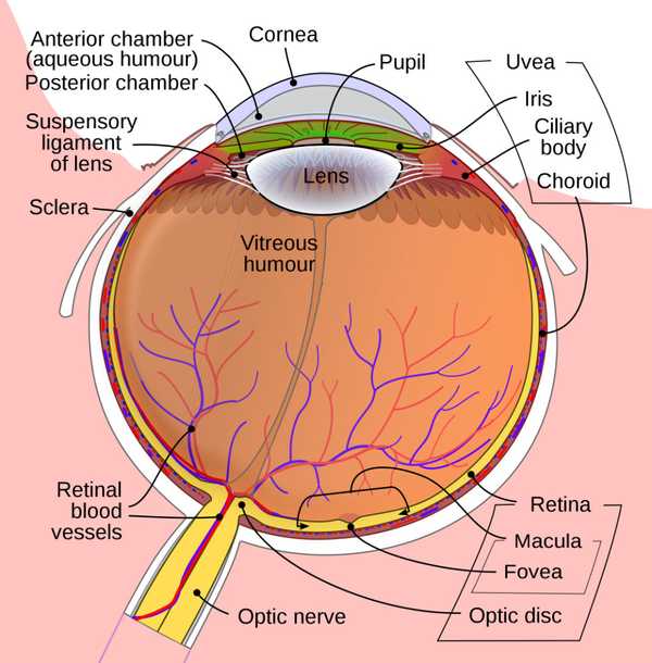
After passing through the cornea, light enters the anterior and posterior chambers of the eye. These chambers are separated by the iris, the colored part of the eye. A water-like liquid called aqueous humour fills these chambers and bathes the iris and the lens with plenty of nutrients.
Moving on, we reach the natural lens within the eye.
The Natural Lens
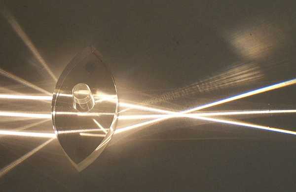
How the lens focuses light; Image by User Fir0002 on en.wikipedia, CC BY-SA 3.0, via Wikimedia Commons
Did you know there is a lens inside your eye? This natural lens often baffles people since when you look at the eye, you can’t see see it! Because of that, many people don’t even know that it exists! But it does exists and serves a pretty critical role.
So where is this mysterious lens? If you look in the pupil of someone, you are starting right at their natural lens. This lens sits right behind the iris or the colored part of the eye. As the name implies (lens), this lens is involved in focusing light. It makes up one third of the focusing power of the eye (with the rest being the cornea). But the lens doesn’t stop there!
One key feature of the lens is that it is soft and flexible and able to change shape. When we are young, this gives us the ability to focus both on things in the distance and things up close; something called accommodation.
How does this lens focus up close?

Seamlessly changing focus; Image by Taibhseoir, CC BY-SA 4.0, via Wikimedia Commons
The whole story of the lens involves another structure: the ciliary body.
The ciliary body is like an inner tube which surrounds the lens. It is a muscle which like all muscles, contracts and relaxes. So how does the ciliary body interact with the lens? The lens is actually connected to the ciliary body through a special “net” called the suspensory ligaments.
The lens, ligaments and ciliary body all dance together and change shape. When we look off in the distance, the ciliary body is relaxed. This pulls the suspensory ligaments tight which flattens the natural lens to focus off in the distance. However, when we look up close, the ciliary body contracts (and moves closer to the lens). This relaxes the tug on the suspensory ligaments pulling the lens. The lens becomes more spherical as a result and the focus changes up close!
And despite all of this changing shape and movement, we still get great distance and up close vision. All in a matter of split second.
But often we don’t appreciate the accommodation of this lens when we are young. As we get older and older, however, we learn more about what this lens has been doing for us all these years. Progressively over time, this lens loses its flexibility. We notice this in our forties when this lens can no longer focus up close for us anymore. After this point, this lens also gets cloudy and starts to impair vision. What is this cloudy lens called? A cataract!
Refractive lens exchange and cataract surgery involve removing this natural lens and replacing it with an artificial lens.
Beyond The Lens
Just a few more stops on our tour: The vitreous humour and the retina. Like the aqueous humour, the jelly-like vitreous humour is a source of nutrients but also provides support to the eye. The vitreous is most notable for being the source of most floaters within the eye.
What Is The Retina?
Finally we reach the retina. Another cool and complex part of the eye.
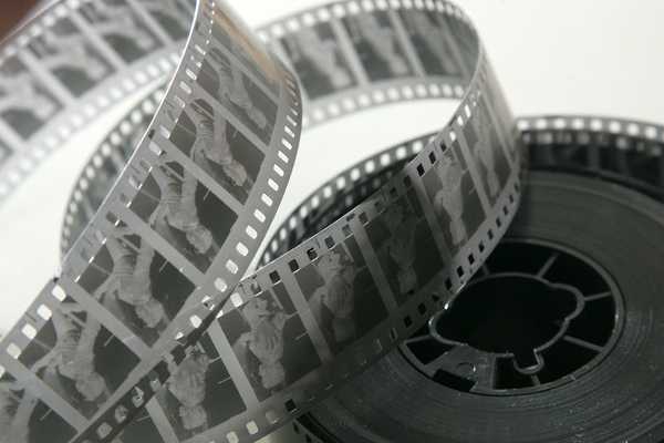
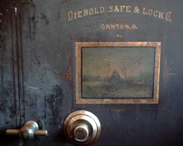
The retina is like camera film; Image by Runner1616 / CC BY-SA
Located at the very back of the eye, the retina is responsible for capturing light. Not unlike the way film from a camera captures pictures. All the light that passes through the cornea and the lens is focused right on the retina. Specialized cells in the retina called rods and cones take these light waves and convert these light waves into signals that the brain can understand. These signals then travel from the retina through a large nerve called the optic nerve out of the eye and to the brain. The brain takes this information to form images and thus our vision!
The center part of the retina is called the macula. The macula has the greatest contribution to how sharp we see things. If you look directly at an object, that object is being projected onto your macula. This is why disorders that affect the macula, such as macular degeneration or retinal detachments that detach the macula, cause so many problems with vision. Having a healthy retina is important to ensure healthy vision.
Amazing that such a thin structure can be so complex!
Also check out Anatomy of the Eye Made Easy on EyeMountain.com to learn more
Related Articles
Please note: The general information provided on the Website is for informational purposes only and is not professional medical advice, diagnosis, treatment, or care, nor is it intended to be a substitute therefore. See the Disclaimer and Terms of Use for more information